EnzyLight™ ADP Assay Kit
Application
- For rapid, quantitative, bioluminescent determination of ADP concentration and evaluation of drug effects on ADP metabolism.
Key Features
- Safe. Non-radioactive assay.
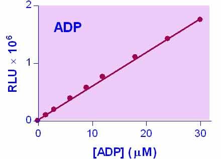
- Sensitive and accurate. As low as 0.02 µM ADP can be quantified.
- Homogeneous and convenient. “Mix-incubate-measure” type assay. No wash and reagent transfer steps are involved.
- Robust and amenable to HTS: Z factors of 0.5 and above are routinely observed in 96-well and 384-well plates. Can be readily automated on HTS liquid handling systems for processing thousands of samples per day.
Method
- Luminescence
Samples
- Cells etc
Species
- All
Procedure
- 20 min
Size
- 100 tests
Detection Limit
- 0.02 µM
Shelf Life
- 12 months
More Details
BioAssay Systems’ EnzyLight™ ADP Assay Kit provides a rapid method to measure ADP levels. The assay involves two steps. In the first step, the working reagent lyses cells to release ATP and ADP. In the presence of luciferase, ATP immediately reacts with the Substrate D-luciferin to produce light. The light intensity is a direct measure of intracellular ATP concentration. In the second step, the ADP is converted to ATP through an enzyme reaction. This newly formed ATP then reacts with the D-luciferin as in the first step. The second light intensity measured represents the total ADP and ATP concentration in the sample. This non-radioactive, homogeneous cell-based assay is performed in microplates. The reagent is compatible with all culture media and with all liquid handling systems for high-throughput screening applications in 96-well and 384-well plates.As we know, generally ADP Enzyme was made by myokinase. Then, how the ADP Enzyme as a kit component made?
Our assay does not use adenylate kinase (aka as myokinase), but pyruvate kinase.
Zhu, J et al. (2020). Altered energy metabolism during early optic nerve crush injury: Implications of warburg-like aerobic glycolysis in facilitating retinal ganglion cell survival. Neuroscience Bulletin, 36(7): 761-777. Assay: ADP in mouse optical tissue.
Su, KH et al. (2019). Heat shock factor 1 is a direct antagonist of amp-activated protein kinase. Molecular Cell, 76(4): 546-561.e8. Assay: ADP in human cells.
Yokoyama, T et al. (2020). Structural and thermodynamic analyses of interactions between death-associated protein kinase 1 and anthraquinones. Acta Crystallographica Section D Structural Biology, 76(5): 438-446. Assay: ADP in human recombinant kinase.
Sun, Shan, et al (2017). Cryo-EM structures of the ATP-bound Vps4 E233Q hexamer and its complex with Vta1 at near-atomic resolution. Nature communications 8: 16064. Assay: ADP in yeast cells.
Ma, Zhenguo, et al (2016). Glyoxylate cycle and metabolism of organic acids in the scutellum of barley seeds during germination. Plant Science 248: 37-44. Assay: ADP in barley seeds.
Ma, Zhenguo, et al (2016). Nitric oxide and reactive oxygen species mediate metabolic changes in barley seed embryo during germination. Frontiers in plant science 7: 138. Assay: ADP in barley seeds.
Cynthia C, Daniel S (2014). Hepatic Lipase Release is Inhibited by a Purinergic Induction of Autophagy. Cell Physiol Biochem 2014;33:883-894. Assay: ADP in human cell.
Cao X, Li LP et al(2013). Astrocytic Adenosine 5′-Triphosphate Release Regulates the Proliferation of Neural Stem Cells in the Adult Hippocampus. Stem Cells. 31(8):1633-43. Assay: ADP in human cells.
Helms CC1, Marvel M et al (2013). Mechanisms of hemolysis-associated platelet activatin. J Thromb Haemost. 11(12):2148-54. Assay: ADP in rodent red blood cell.
Narain, NR (2011). Methods For Treatment Of Metabolic Disorders Using Epimetabolic Shifters, Multidimensional Intracellular Molecules, Or Environmental Influencers. US 2011/0020312 Al. Assay: ADP in mouse cell.
Ponnusamy M, et al (2011). P2X7 receptors mediate deleterious renal epithelial-fibroblast cross talk. Am J Physiol Renal Physiol. 300(1):F62-70. Assay: ADP in human epithelial cells.
Vacirca D, et al (2011). Anti-ATP Synthase Autoantibodies from Patients with Alzheimer’s Disease Reduce Extracellular HDL Level. J Alzheimers Dis. 26(3):441-5. Assay: ADP in human neuronal cells.
Belleannee C, et al (2010). Role of purinergic signaling pathways in V-ATPase recruitment to apical membrane of acidifying epididymal clear cells. Am J Physiol Cell Physiol. 298(4):C817-30. Assay: ADP in human epididymal cells.
Saito A, Castilho RF (2010). Inhibitory effects of adenine nucleotides on brain mitochondrial permeability transition Neurochem Res.35(11):1667-74. Assay: ADP in mouse brain tissue.
To find more recent publications, please click here.
If you or your labs do not have the equipment or scientists necessary to run this assay, BioAssay Systems can perform the service for you.
– Fast turnaround
– Quality data
– Low cost
Please email or call 1-510-782-9988 x 2 to discuss your projects.

$419.00
For bulk quote or custom reagents, please email or call 1-510-782-9988 x 1.
Orders are shipped the same day if placed by 2pm PST
Shipping: On Ice
Carrier: Fedex
Delivery: 1-2 days (US), 3-6 days (Intl)
Storage: -20°C upon receipt
Related Products
You may also like…
| Name | SKU | Price | Buy |
|---|---|---|---|
| EnzyLight™ ATP Assay Kit | EATP-100 |
$419.00 |
|
| EnzyLight™ ADP/ATP Ratio Assay Kit | ELDT-100 |
$459.00 |
|
| EnzyChrom™ ADP Assay Kit | E2ADP-100 |
$479.00 |
|
| QuantiChrom™ ATPase Assay Kit | DATG-200 |
$469.00 |
Why BioAssay Systems
Quality and User-friendly • Expert Technical Support • Competitive Prices • Expansive Catalogue • Trusted Globally
