NFκB Phosphorylation Status Screening Service
Summary
Using our EnzyFluo™ NFκB Phosphorylation Assay Kit, we screen for modulators that increase or decrease NFκB phosphorylation status in cells. We measure the NFκB phosphorylation kinetics, EC50, and IC50 of cells treated with modulators. Testing can be run on a wide variety of cell lines.
Species Reactivity
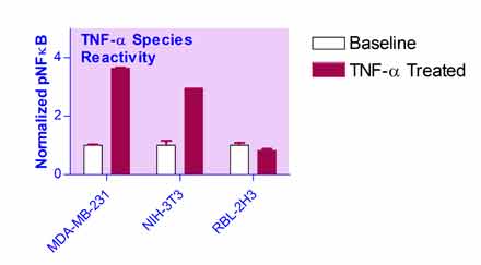
We first determine the test compound’s reactivity with different species. In this example, the species tested were human (MDA-MB-231), mouse (NIH-3T3), and rat (RBL-2H3). Here we use Tumor necrosis factor alpha (TNF-α) as an example test compound. In a sterile, black 96-well plate, MDA-MB-231, NIH-3T3, and RBL-2H3 cells were plated at a cell density of 20,000 cells per well and allowed to adhere overnight. The next day, the culture medium was removed and replaced with medium dosed with 10 ng/mL TNF-α or no TNF-α for the baseline controls. The dosed culture medium was prepared from a stock solution of 10,000 ng/mL TNF-α in dH2O. Triplicate wells were incubated for 10 minutes with the TNF-α or control medium. The pNFκB and total protein was then quantified using the NFκB Phosphorylation Assay Kit (EFNKB-100). The pNFκB values were normalized to the total protein content of the sample.
The relative increase in pNFκB for the cells treated with TNF-α was 3.64:1, 2.96:1, and 0.83:1 for MDA-MB-231, NIH-3T3, and RBL-2H3 respectively. The cell line MDA-MB-231 showed the greatest reactivity to the test compound TNF-α and was chosen as the cell line for the next experiments.

Phosphorylation Titration
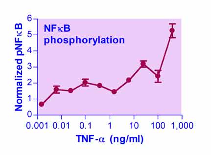
To determine the time course of NFκB phosphorylation, first a titration was run on the concentration of the test compound; here we use TNF-α as an example. In a sterile, black 96-well plate, MDA-MB-231 cells were plated at a cell density of 20,000 cells per well and allowed to adhere overnight. The next day, the culture medium was removed and replaced with medium dosed with TNF-α at the following concentrations: 0.0E+0, 3.8E-4, 1.5E-3, 6.1E-3, 2.4E-2, 9.8E-2, 3.9E-1, 1.6E+0, 6.3E+0, 2.5E+1, 1.0E+2, and 4.0E+2 ng/mL. The dosed culture medium was prepared from a stock solution of 10,000 ng/mL TNF-α in dH2O. Triplicate wells were incubated for 20 minutes with the TNF-α. The pNFκB and total protein was then quantified using the NFκB Phosphorylation Assay Kit (ENFKB-100). The pNFκB values were normalized to the total protein content of the sample.

Phosphorylation Kinetics
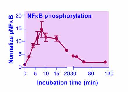
After the titration on the test compound concentration, we measure the NFκB phosphorylation kinetics for a specific dosage. TNF-α will continue to serve as our example here; we measure the NFκB phosphorylation kinetics for TNF-α at a concentration of 100 ng/mL. In a sterile, black 96-well plate, PANC-1 cells were plated at a cell density of 20,000 cells per well and allowed to adhere overnight. The next day, the culture medium was removed and replaced with 100 ng/mL TNF-α dosed medium. The dosed culture medium was prepared from a stock solution of 10,000 ng/mL TNF-α in dH2O. Triplicate wells were incubated for 0, 2, 4, 6, 8, 10, 15, 20, 30, 40, 60, and 120 minutes with the TNF-&alpha. The pNFκB and total protein was then quantified using the NFκB Phosphorylation Assay Kit (ENFKB-100). The pNFκB values were normalized to the total protein content of the sample. The 0 minutes (no TNF-α incubation) served as the untreated control for basal NFκB phosphorylation.

Please email or call 1-510-782-9988 x 2 to request assay service.
Customer Testimonials
"The guys at BioAssay Systems know their science! I had to develop a bioassay for our company and Robert was very quick to respond to keep us up to date. They are knowledgeable and very resourceful. They directed us to one of the bulk suppliers that could give us a really good deal on our active ingredient. They keep very detailed records for our research needs. I would recommend working with BioAssay Systems on your assay development projects!"

"It is a pleasure working together with BioAssay Systems. I brought samples to their Hayward location and received the results in less than 24 hr. The discussion with Robert Z was courteous, open, and with a depth of knowledge. Robert has a PhD from Stanford. Also, Frank Huang, the CEO, had sufficient scientific curiosity to try out our cleanser and provide feedback! This is beyond the call of customer relations. BioAssay Systems is a company with a lot of expertise and a friendly, responsive approach to problem solving. I will enthusiastically continue to work with them to carry out our research."

"I would like to show my appreciation to the Bioassay Systems service team. We are developing an assay to measure the change in lactate concentration of single cell samples using fluorescent microscopy. The BioAssay Systems team was quite helpful in assisting us to optimize their fluorescent L-Lactate assay kit for our unique experimental set-up. The BioAssay Systems team is always friendly and knowledgeable. We know that we can rely on them for fast and professional service."

"While developing biodegradable coatings for Drug Eluting Stents I worked with Bob to develop an accurate, precise and sensitive assay for determining the amount of polymer remaining in tissue. It was a pleasure to work with Bob and his team, they are very thorough, they understand the science very well and they always deliver on time and at the right price. I would highly recommend anyone using BioAssay Systems services in the future."

